Case report. Goblet Cell Carcinoid of the Appendix. A 52 years old male with acute appendicitis
CASE REPORT: Goblet Cell Carcinoid of the Appendix. A 52 years old male with acute appendicitis.
Rafael Trujillo Vilchez, Diego Pérez Martín, Julia Alcaide García, Rafael Funez Liebana (a), Fco Javier Moya Donoso (b) and Antonio Rueda Dominguez (c).
Oncology Department. Hospital of the Costa del Sol. Marbella. Spain
a. Department of Pathology. Hospital of the Costa del Sol. Marbella. Spain
b. Department of Surgery. Hospital of the Costa del Sol. Marbella. Spain
c. Chief Head Department of Oncology . Hospital of the Costa del Sol. Marbella. Spain
Goblet cell carcinoid, also variably known as adenocarcinoid, mucinous carcinoid, and crypt cell carcinoma, is a rare neoplasm with distinct histological and clinical features. We review the management of goblet cell carcinoid of the appendix using an illustrative case report.
Key Words: Goblet cell carcinoid. Adenocarcinoid. Small-bowel neoplasms. Appendix.
Introduction
Goblet cell carcinoid, also variably known as adenocarcinoid, mucinous carcinoid, and crypt cell carcinoma, is a rare neoplasm with distinct histological and clinical features. It has histological features of carcinoid as well as adenocarcinoma and was initially described in a large series by Subbuswamy et al (1) in 1974. It is believed to arise from crypt base stem cells (2) Generally it stains positive for synaptophysin, neuron-specific enolase, cytokeratin,chromogranin, and bioamines, which are characteristic of carcinoid lineage (3,4). However, it does not secrete the active hormones, i.e., serotonin and its by products, typically seen in carcinoid, at least not to the extent detectable in systemic circulation or enough to produce carcinoid symptoms. Goblet cell carcinoid produces mucin, a feature consistent with adenocarcinoma cell line (3,5)
We review the management of goblet cell carcinoid of the appendix using an illustrative case history.
Case reports
A 52 years old male was admitted in our Hospital with a four days history of right iliac fossa abdominal pain associated with diarrhoea. On examination he was apyrexic with rebound tenderness in the right iliac fossa. Blood investigations revealed a high white cell count Abdominal sonography was performed which showed thickened appendix compatible with acute appendicitis. An urgent appendicectomy was performance, which confirmed an acutely inflamed appendix.
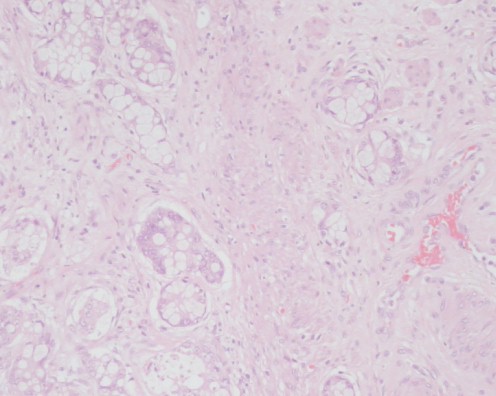
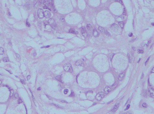
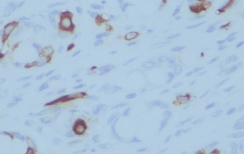
Histopathology confirmed acute appendicitis. In addition, there was a tumour at the tip of the appendix with clear margins of 6 mm, composed of small glandular acini and individual cells with eosinophilic and focally granular cytoplasm (figure 1 and 2). It stains positive for chromogranin (figure 3). Overall the histological features were those of a goblet cell carcinoid tumour of the appendix with co-existing acute appendicitis.


Post-operatively an urinary levels of 5-hydroxyindoleacetic acid (5-HIAA) were within normal limits. An octreotide radioisotope scan did not reveal any metastatic disease. It was felt that in view of clear margins of only 6 mm, to performance a right hemicolectomy was justified to decrease his risk of delayed local and distant recurrence, and to study the regional lymph nodes. He proceeded to a laparoscopic right hemicolectomy, the pathology of which did not reveal any residual disease and at eight month follow up he remains disease free.
Discussion
The diagnosis of goblet cell carcinoid of the appendix is essentially made on histological examination after surgery, which is one of the clinical hallmarks of appendiceal carcinoids. This neoplasm has also been described as an adenocarcinoid, crypt cell carcinoma and goblet cell carcinoma. The preferred term, goblet cell carcinoma, was first coined in 1974 by Subbuswamy et al (1) as the nomenclature implies, these tumours possess morphological features suggestive of both carcinoid and glandular differentiation.
Reports from the medical literature on goblet cell carcinoid are sparse; however, the general consensus is that the clinical behaviour of goblet cell carcinoid is intermediate between carcinoid and adenocarcinoma (6–8). It is a more aggressive neoplasm compared with appendiceal carcinoid but is more indolent compared with adenocarcinoma (9)
They are characterized histologically by wide invasion of the mesoappendix, and clinically by delayed local recurrence and distant metastases. As the vast majority of these tumours are detected postoperatively, management centres on the need for re-operation as a right hemicolectomy is often curative (10). In patients with goblet cell carcinoids
relative indications that may favour further surgery are:
(A) Angioinvasion as an isolated finding
(B) Tumours with clear margins less than 2 cm
(C) Mucin-producing tumours. (11)
In our case the decision to performance a right hemicolectomy was justified to decrease his risk of delayed local and distant recurrence, and to study the regional lymph in view of clear margins of only 6 mm.
Conclusions
Goblet cell carcinoid is a rare malignancy that arises mostly from the appendix but can also occur in other parts of the small bowel. Its prognosis is intermediate between classic carcinoid and adenocarcinoma of the appendix, with mean survival of 47 months and an overall 5-year survival of 45%. Goblet cell carcinoid has a propensity for peritoneal seeding that leads to peritoneal carcinomatosis. Careful diagnosis and management of goblet cell carcinoid tumours will hopefully improve prognosis in patients with this rare entity.
Conflict of interest
The authors have no conflict of interest
References
1.Subbuswamy SG,Gibbs NM,Ross CF,Morson BC. Goblet cell carcinoid of the appendix. Cancer 1974; 34:338–44.
2. Warner TF,Seo IS. Goblet cell carcinoid of appendix: ultrastructural
features and histogenetic aspects. Cancer 1979; 44:1700–6.
3. Hofler H,Kloppel G,Heitz PU. Combined production of mucus,amines and peptides by goblet-cell carcinoids of the appendix and ileum. Pathol Res Pract 1984; 178:555–61.
4. Burke AP,Sobin LH,Federspiel BH,Shekitka KM. Appendiceal
carcinoids: correlation of histology and immunohistochemistry. Mod Pathol 1989; 2:630–7.
5. Chen V,Qizilbash AH. Goblet cell carcinoid tumor of the appendix. Report of five cases and review of the literature. Arch Pathol Lab Med 1979; 103:180–2.
6.Berardi RS,Lee SS,Chen HP. Goblet cell carcinoids of the appendix. Surg Gynecol Obstet 1988; 167:81–6.
7. Anderson NH,Somervil le JE,Johnsto n CF,Hayes DM,Buchanan KD,Sloan JM. Appendiceal goblet cell carcinoids: a clinicopathological and immunohistochemical study. Histopathology 1991; 18:61–5.
8. Butler JA,Houshia r A,Lin F, Wilson SE. Goblet cell carcinoid of the appendix. Am J Surg 1994; 168:685–7
9. McCusker ME,Cote TR,Clegg LX,Sobin LH. Primary malignant neoplasms of the appendix: a population-based study from the surveillance,epidemiology and end-results program,1973-1998. Cancer 2002; 94:3307–12.
10. Kanthan R, Saxena A, Kanthan SC. Goblet cell carcinoids of the appendix: immunophenotype and ultrastructural study. Arch Pathol Lab Med 2001; 125: 386-390.
11. Goede AC, Caplin ME, Winslet MC. Carcinoid tumour of the appendix. Br J Surg 2003; 90: 1317-1322.