Ovarian fibroid in pregnancy. Case presentation.1
Ovarian fibroid in pregnancy. Case presentation
P. Baffoe, O. Fofie and B. Ferreiro. Department of Obstetrics and Gynaecology, Bolgatanga regional hospital, P.O.Box 26, Bolgatanga, Ghana.
Summary
Ovarian tumor is one of the most common conditions that pose a challenge to a gynaecologist especially when it’s solid in nature. In general about 80% of all ovarian tumors are benign with favorable prognosis.
The other 20% are malignant. Benign ovarian tumors are often diagnosed accidentally if they are not large but when they become large patients could present with abdominal discomfort, pains, constipation etc. due to compression on other pelvic and abdominal organs.
Ovarian fibroid is a solid benign ovarian tumor with favorable prognosis. It’s not very frequent. This is a simple connective tissue tumor originating from the mesenchymal and can easily be confused with thecoma. The association of ovarian fibroid and uneventful pregnancy is considered as an extreme rarity. Very few cases of such association have been published in some prominent literature. We are motivated to present this case due to its rarity and the unpredictability of its presenting forms.
Key words: Ovarian fibroid
Introduction
Ovarian fibroid is a solid benign ovarian tumor with good prognosis. The risk of malignancy is less than 1% (degeneration into fibrosarcoma). These tumors are generally asymptomatic thus making diagnosis very difficult when they are of smaller dimension. In most of the cases it’s discovered accidentally during laparotomy or pelvic ultrasound. They are generally slow growing tumors, however, ovarian fibroids may become very big with time and pressure symptoms ensue due to compression on pelvic and abdominal organs.
These tumors are generally more frequent at the post-menopausal age but some studies have not found any difference between pre and post menopausal women as far as incidence is concerned. In nine years studies, Wai Leung and Pong Mo Yuen of Prince of Wales Hospital, University of Hong Kong, China, found that out of twenty three (23) patients who were diagnosed of ovarian fibroid, representing just 1% of all benign ovarian tumors, the average age of these patients was 45 years and 47.8% were in post menopausal age and the rest were premenopausal women. The average size of these tumors was 13cm. and all were unilateral tumors.
Macroscopically, ovarian fibroid are of the same appearance as uterine fibroid but microscopically, cystic degeneration could be seen on the cortical surface, the cut surface is whorled and trabecular in appearance. There are spindle cells which are composed of fibroblastic ovarian cells with stromal appearance. There may be some papillary projections on the cortical surface.
Due to the rarity of the association of these tumors with uneventful pregnancy we have decided to present this case that was managed at the Department of Obs. & Gynae. Bolgatanga Regional Hospital, Ghana in December 2007.
Case summary
This patient was 32 years old referred from the Builsa district hospital on the 3/12/2007 at 3.00am due to persistence of huge abdominal mass and abdominal pains after delivery. Patient had her first menstruation at the age of 13, Gravida 2 Para 1. Past medical history was unremarkable. On examination: Pallor+, no fever, pulse 88/min. BP 110/70mmHg. Respiratory and cardiovascular systems were apparently normal.
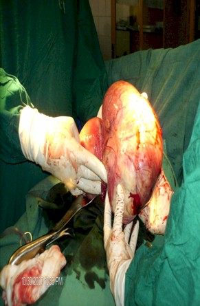
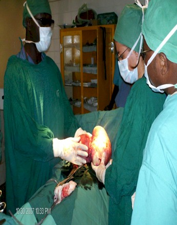
Abdomen was distended and tender on palpation with a huge mass arising from pelvis, firm, hard very tender when mobilized. Vaginal examination revealed closed cervix, lochia rubra. Uterus well contracted and retracted. In ultrasound studies, uterus was increased in size in accordance with immediate post-delivery. Right ovary was normal. Left ovary was grossly enlarged with an echogenic mass measuring 16cm x 10cm x 8cm. Hemoglobin level 7.3g/dl and blood group B positive. 2 units of B positive blood were transfused and patient was prepared for laparotomy due to a complicated ovarian tumor. General Anaesthesia was applied.

